
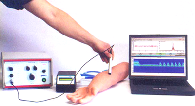

Description
Basics
Doppler Sonography uses the doppler effect to assess whether structures (usually blood) are moving towards or away from the ultrasonic probe,and its relative velocity. By calculating the frequency shift of a paticular sample volume, for example a jet of blood flow over a heart valve, speed and direction of sample volume can be determined and visualised. Doppler frequency shift is the difference in ultrasound frequency between transmitted and received echoes, the echoes frequency minus the transmitted frequency. The doppler frequency is proportional to the blood flow velocity. Doppler sonography is used for cardiovascular study (sonography of the vasculature system and heart) and reverse blood flow in the liver.
Procedure
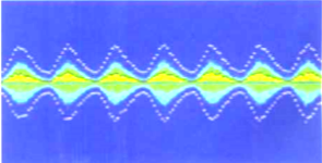
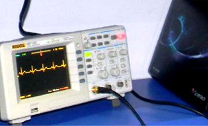
The pump is switched on and the speed is adjusted in a middle range (ca. 4000 min -1). The mode is GK (continuously, venous). With the doppler probe and coupling gel the arm model is scanned for a vessel with a significant audio signal. The flow in the spectral image is analysed for negative and positive components . The probe direction is then switch by 180º. Then the vessel is scanned for changes in the spectral image and the differences between the image of healthy vessel and the stenosis will be characterised.The pump is switched to P1 and P2 Mode (pulsatil ) the image are analysed and pulse rate is determine.
Results
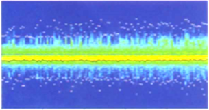
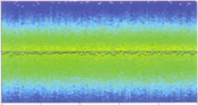
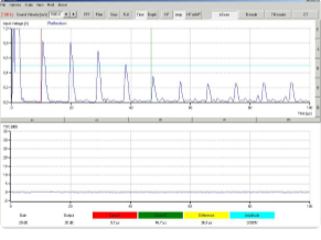
Figure (1) Shows a continuously (venous) flow with a mean doppler shift of ca.-700Hz . The minus in the doppler shift means flow away from the probe. Figure(3) Shows the doppler spectral fingure of a stenosis. The differences to a normal (healthy) fingure like Figure 1 are:
- A local increase of the maximum doppler shift (maximum flow velocity).
- A decrease of mean frequency and a broadening of the spectra.
- An increase of reflux phenomenon (negative and positive parts of the spectra).


")


Reviews
There are no reviews yet.