


Description
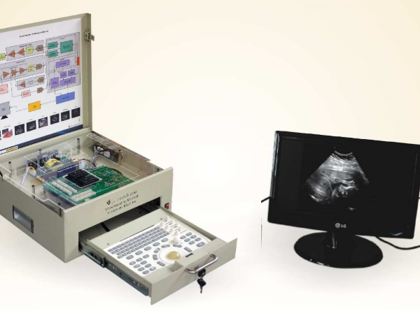
Scientech 2364 Working of Medical Ultrasound Machine is designed in such a manner that it provides full technical information of both medical and electronic parts. Students can learn different techniques for the analysis of ultrasound imaging.
Features
- Easy to use and specially design for educational purpose
- Calculation and analysis of images with the help of software
- Direct interface with printer
- Direct interface with LCD with the help of VGA connector
- Additional probe interface (optional)
- USB and Serial port interfacing
- DICOM interfacing (optional)
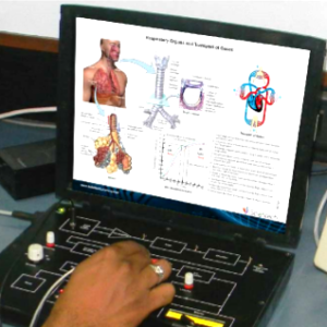
- Software package for heart analysis and calculations
Scope of Learning
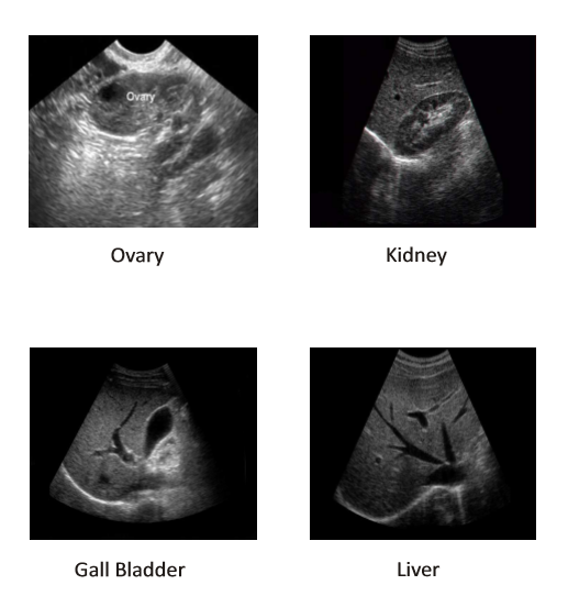
Analysis of:
- Liver and Gall bladder
- IVC and Hepatic Portal System
- Pancreas
- Spleen and Spleenic ducts.
- Kidney (Right & Left)
- Uterus
- Ovary (Right & Left)
- Urinary bladder
- Prostate gland
- Aorta and IVC
- Heart in Parasternal Long Axis (PLAX)
- Heart in Parasternal Short Axis (PSAX)
- Heart in Subcostal view position
- Heart in Suprasternal view
- Mitral and Aortic valve in M mode of echocardiography
Standard Configuration :
- Main unit
- 3.5 MHz Convex probe (80e R60)
- Cine loop system (256 F)
- Color image output (16 kinds)
Optional :
- 6.5 MHz Trans – vaginal probes
- 7.5 MHz Linear probe
- Dual probe socket
- USB
- Linear Probe
- Convex Probe
- Trans-Vaginal Probe
Technical Specifications
- Probe : 2.0 – 5.0 MHz multi-frequency convex probe
- Display : B, B+B, 4B, B+M, M
- Scanning depth : = 80 mm
- Gray scales : 256
Resolution :
- Lateral resolution :
= 2 mm (depth = 80 mm);
= 3 mm (80 < depth = 130 mm) Axial := 1 mm (depth = 80 mm);
= 2 mm (80< depth = 130mm);
- Dead zone : = 3mm
- Geometry position precision: lateral = 5% Axial = 5%
- Scanning line : 512/frame, frequency : 30 frames / second
- Focusing : 4 focuses (dynamic variable aperture)
- STC adjustment : 8 segment TGC
- Image processing : Changeable aperture; dynamic filter, Dynamic frequency scanning, L/R, UP/DOWN, edge enhancement; multistage electronic focus
- Dynamic range : 64~96 dB adjustable
- Zoom in : ×0.8, ×1.0, ×1.2, ×1.5, ×1.8 and × 2.0
- PIP : Display PIP can show the image clearer
- Body marks : 16
- Measurement : Distance, circuit, size, volume, BPD, GS, CRL, FL, AC, HC
- Connectors : VGA, PAL – D
- Monitor : 10″ B / W monitor (SVGA noninterlaced)
- Pseudo color : Connection with normal monitor is available
- Cine loop : 256 frames, automatic or manual
- Permanent Image memory : 128 frames
- Power : AC 220/110V, 50/60 Hz
- G/W : 11 KGs
- N/W : 7.5 KGs
- Packing size : 480 × 380 × 410 mm Carton packing
Included Accessories :
- Transducer Probe : 1 no. (3.5 MHz Convex)
- Ground Cable : 1 no. Fuse (1A) : 2 nos.
- Ultrasound gel : 1 no
- Display Monitor (TFT) : 1 no
- Mains Power Cord : 1 no
- USB Cable A-type : 1 no (Male to Male)
- Tissue Paper packet : 1 no


")


Reviews
There are no reviews yet.